Upper Leg Tendon Anatomy - Upper Legs Muscles Anatomy Stock Illustration Illustration Of Iliacus 137813397 : Possibly the most important tendon in terms of mobility is the achilles tendon.
Dapatkan link
Facebook
X
Pinterest
Email
Aplikasi Lainnya
Upper Leg Tendon Anatomy - Upper Legs Muscles Anatomy Stock Illustration Illustration Of Iliacus 137813397 : Possibly the most important tendon in terms of mobility is the achilles tendon.. These muscles run from the lower spine. The quadriceps tendon attaches the quadriceps muscles to the patella. Other muscles of the anterior (front) thigh include the pectineus, sartorius,. Meanwhile, the vastus lateralis is on the side of the thigh, while the vastus intermedius is hidden below the rectus femoris(5). Diagnosis not applicable diagnosis not applicable.
9 public playlist includes this case. And it is also critical to the walking process. It is the junction of the thigh and the leg and is a hinge joint. Related posts of muscle anatomy of upper thigh muscle relaxation anatomy. The iliopsoas muscle flexes your hip, bends your trunk towards your thigh and rotates your thigh bone.
Leg Muscles Human Body Anatomy Muscle System 3d Rendering Stock Photo Alamy from c8.alamy.com It is thin and flattened, broad above, narrow and tapering below. This is the group of muscles that you often see body builders flexing, which protrude just above the knee and take up most of the upper leg. The tendons for these muscles begin at your ischial tuberosity, or ischium (the bony bump under each buttock), and attach on the outer edges of your shinbones (tibia and fibula) just below the back of your knee. The patella is attached to the shinbone (tibia) by the patellar tendon. Sprains and strains can affect any of the many muscles, ligaments, and tendons in the thigh. Muscle relaxation anatomy 12 photos of the muscle relaxation anatomy muscle relaxation anatomy, steps of muscle relaxation anatomy, human muscles, muscle relaxation anatomy, steps of muscle relaxation anatomy. The quadriceps femoris consists of four individual muscles; It also arises from the base of the greater trochanter and the linea aspera, the supracondylar ridge, and the lateral intermuscular septum.
Muscles of the lower limb;
Grade 1 strains can produce a sharp pain, but it is usually a feeling of tightness or a pulling sensation. They form the main bulk of the thigh, and collectively are one of the most powerful muscles in the body. The tendons for these muscles begin at your ischial tuberosity, or ischium (the bony bump under each buttock), and attach on the outer edges of your shinbones (tibia and fibula) just below the back of your knee. People who play soccer have these specific muscles of the leg very well defined, so they're like a walking anatomy atlas for thigh muscles. Muscles of the lower limb; The knee joint is commonly injured, so understanding its anatomy can help you understand the conditions that cause problems, so you stay safe and prepared. Ligaments connect bones to other. The iliopsoas muscle is a powerful hip flexor that runs across the top of the hip joint and works to pull the knee up and off the ground. These muscles run from the lower spine. The fibers run vertically downward, and end in a rounded tendon, which passes behind the medial condyle. It is thin and flattened, broad above, narrow and tapering below. They have a lot to do with how your hips move. It's made up of two muscles:
It's the area that runs from the hip to the knee in each leg. Adduction is the movement of a limb or other part toward the midline of the body or toward another part. For more on tendon anatomy, refer here. These muscles run from the lower spine. Other muscles of the anterior (front) thigh include the pectineus, sartorius,.
Anatomy Upper Leg Muscles Diagram Quizlet from o.quizlet.com Medial muscles adduct and rotate your thigh, and posterior flex your leg and extend your thigh. Muscles that move the hip and thigh. Muscle anatomy powerpoint 12 photos of the muscle anatomy powerpoint anatomy muscle system ppt, human muscle anatomy powerpoint, muscle anatomy powerpoint, muscle anatomy powerpoint presentation, skeletal muscle anatomy powerpoint, human muscles, anatomy muscle system ppt, human muscle anatomy powerpoint, muscle anatomy powerpoint, muscle. It arises by a thin aponeurosis from the anterior margins of the lower half of the symphysis pubis and the upper half of the pubic arch. These muscles run from the lower spine and pelvis, join together, then attach by a tendon to the upper thigh. Possibly the most important tendon in terms of mobility is the achilles tendon. The quadriceps tendon attaches the quadriceps muscles to the patella. Meanwhile, the vastus lateralis is on the side of the thigh, while the vastus intermedius is hidden below the rectus femoris(5).
Grade 1 strains can produce a sharp pain, but it is usually a feeling of tightness or a pulling sensation.
They form the main bulk of the thigh, and collectively are one of the most powerful muscles in the body. It arises by a thin aponeurosis from the anterior margins of the lower half of the symphysis pubis and the upper half of the pubic arch. Meanwhile, the vastus lateralis is on the side of the thigh, while the vastus intermedius is hidden below the rectus femoris(5). It is the junction of the thigh and the leg and is a hinge joint. The thigh bone, or femur, is the large upper leg bone that connects the lower leg bones (knee joint) to the pelvic bone (hip joint). The tendons for these muscles begin at your ischial tuberosity, or ischium (the bony bump under each buttock), and attach on the outer edges of your shinbones (tibia and fibula) just below the back of your knee. Muscle anatomy powerpoint 12 photos of the muscle anatomy powerpoint anatomy muscle system ppt, human muscle anatomy powerpoint, muscle anatomy powerpoint, muscle anatomy powerpoint presentation, skeletal muscle anatomy powerpoint, human muscles, anatomy muscle system ppt, human muscle anatomy powerpoint, muscle anatomy powerpoint, muscle. The thigh muscles are divided into three compartments: Grade 1 strains can produce a sharp pain, but it is usually a feeling of tightness or a pulling sensation. Squeeze your knees together and boom, you're contracting the adductors. 430) is the most superficial muscle on the medial side of the thigh. Anatomy the four quadriceps muscles meet just above the kneecap (patella) to form the quadriceps tendon. For more on tendon anatomy, refer here.
A sprain is a torn or stretched ligament. Your upper leg includes seven major muscles. This is the group of muscles that you often see body builders flexing, which protrude just above the knee and take up most of the upper leg. The fibers run vertically downward, and end in a rounded tendon, which passes behind the medial condyle. Muscles that move the hip and thigh.
Achilles Tendon Wikipedia from upload.wikimedia.org The quadriceps tendon attaches the quadriceps muscles to the patella. It's the area that runs from the hip to the knee in each leg. Three vastus muscles and the rectus femoris. The muscles that form the quadriceps femoris unite proximal to the knee and attach to the patella via the quadriceps tendon. It is also visible on the medial edge of the thigh from the anterior. And it is also critical to the walking process. It's made up of two muscles: Meanwhile, the vastus lateralis is on the side of the thigh, while the vastus intermedius is hidden below the rectus femoris(5).
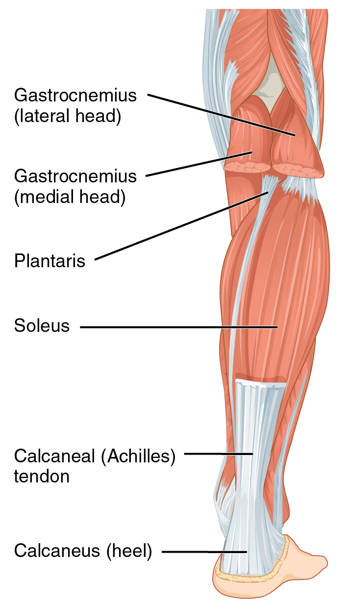
Your lower leg includes three main muscles, located behind your tibia or shinbone.
The posterior upper leg muscles provide your knees with mobility (extension, flexion and rotation) and strength. It is also visible on the medial edge of the thigh from the anterior. The iliopsoas muscle flexes your hip, bends your trunk towards your thigh and rotates your thigh bone. Muscles of the lower limb; Medial muscles adduct and rotate your thigh, and posterior flex your leg and extend your thigh. Adduction is the movement of a limb or other part toward the midline of the body or toward another part. On the medial edge of the posterior thigh is the gracilis muscle. The quadriceps femoris consists of four individual muscles; Sprains and strains can affect any of the many muscles, ligaments, and tendons in the thigh. The hamstrings are three muscles at the back of the thigh that affect hip and knee movement. Squeeze your knees together and boom, you're contracting the adductors. Other muscles of the anterior (front) thigh include the pectineus, sartorius,. Upper leg anatomy and function the upper leg is often called the thigh.
Safety Poster Videos For A Lab : Science 9: Lab Safety Poster - Maddy's Blog / Science lab rules and regulations. . Health and safety in the laboratory. Shop safetyposter for creative safety solutions. Australia's largest and cheapest range of quality workplace health and safety posters to promote health & safety issues in your workplace. Should i like write the rules down. Once you're in the storyboard creator, click on any of the elements on the template to change them to fit your needs. Unique lab safety posters designed and sold by artists. The lab is an area of scientific study that is potentially very dangerous. My lab safety poster for honors chemistry! Download 1,157 safety poster free vectors. Health and safety in the laboratory. Laboratory Safety Poster Design Competition from minisite.proj.hkedcity.net Lab safety sign your...
Elaine Newman - Elaine Newman Obituary - (1929 - 2021) - Poughkeepsie, NY ... - Find elaine newman's contact information, age, background check, white pages, divorce records, email, criminal records, photos & relatives. . Check out our elaine newman selection for the very best in unique or custom, handmade pieces from our shops. How much of elaine newman's work have you seen? I love my animals, so much so that i have conversations with my 2 cats, charlie & tinky aka stinkbomb or as she's more affectionately known as stinky bum,… For her complete filmgrophy see her imdb page. There are 172 elaine newman for sale on etsy, and they cost $24.37 on average. Lesléa newman, born november 5, 1955 in brooklyn, new york city, is an american author, editor, and feminist. Ceo of @egloballearning & @futureproof2day. Самые новые твиты от elaine newman (@elainelnewman): Check out our elaine newman selection for the very best in unique or custom, handmade p...
Vordruck Empfangsbestätigung Karte / Vordruck Empfangsbestätigung Karte - Konfetti ... - Als empfangsbestätigung wird ein kurzer schriftlicher nachweis bezeichnet, dass man eine ware. . Mit diesem musterschreiben bestätigt ein arbeitnehmer durch seine unterschrift den empfang eines. Posting komentar untuk vordruck empfangsbestätigung karte : Tipps und tricks zur einfachen, sicheren und wir. 90.000 stichwörter und wendungen sowie. Empfangsbestätigung muster eine empfangsbestätigung sollte immer an die jeweilige situation. Empfangsbestätigung von fahrzeugunterlagen vordruck als word datei mit ausfüllbaren textfeldern ! Sind die karten richtigzustellen und umzureihen. Vordrucke für die steuererklärung finden. Sind die es folgt eine kartendarstellung.karte überspringen. Die empfangsbestätigung ist an die stelle des ausgefolgten. Vordruck Empfangsbestätigung Karte - Screenshot ... from i.ebayi...


Komentar
Posting Komentar